Mary Kelly blood vessels
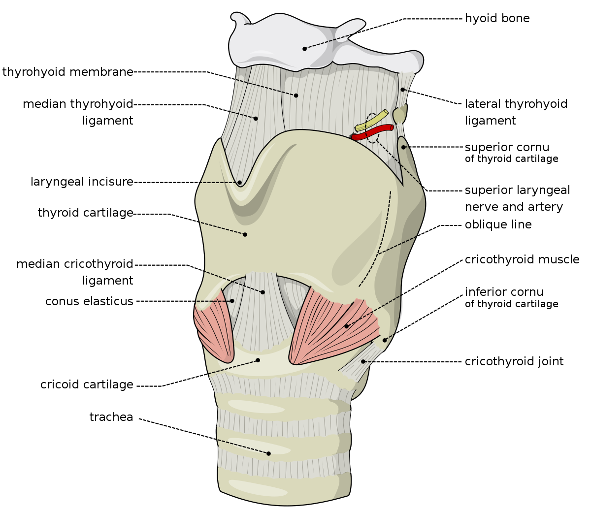
This post is not intended to be comprehensive, but a jumping off point for my buddy Joel. The image contains red circles around vessels that would have to be addressed in accomplishing the described wounds. Dave
Description,"The neck was cut through the skin & other tissues right down to the vertebrae the 5th & 6th being deeply notched. The skin cuts in the front of the neck showed distinct ecchymosis."
description,"The Pericardium was open below & the Heart absent."
description,"The viscera were found in various parts viz: the uterus & Kidneys with one breast under the head, the other breast by the Rt foot, the Liver between the feet, the intestines by the right side & the s pleen by the left side of the body. The flaps removed from the abdomen and thighs were on a table."
Ripper Anatomy Class
Collapse
X
-
I have helpers!
...and oh I am so glad for it! A very big shout out to Joel who is making medically less ignorant, I am sure at some cost to his patience. He is great help in achieving accuracy for me.
...and since I am software challenged as well, an equally large shout out to Jane who is equally generous in explaining the software and even doing the stuff that is clearly beyond my hope to do correctly.
In terms of how the reader should understand this, NO POST on this thread would be possible without their efforts in helping my dumb ass, So while my name is on the post, it is in fact a triad of skill making these images correct and understandable. If the truth were told, I am the weakest link in the damn chain. Dave
Leave a comment:
-
-
-
mary Kelly basics
In understanding Mary's described injuries we need to start from scratch again. Here is an illustration of body cavitations. We need to do this in order to grasp the severity of inflicted injuries.
1) wound description, "The pericardium was open below and the heart absent." This places the killer in the thoracic cavitation (top of diagram).
2) wound description,"The whole of the surface of the abdomen and thighs was removed and the abdominal cavity emptied of its viscera." This places the killer in the abdominal cavitation. (middle of diagram and separated from the aforementioned cavitation by the physical structure of the diaphragm.
3) wound statement, " The viscera were found in various parts viz: the uterus and kidneys with one breast under the head," This places the killer in the lowest cavitation on the diagram.
And so it must be realized that the killer thoroughly explored the interior cavitations of Ms. Kelly. Because there is a physical structure separating two of these cavitations, it can be safely inferred that this was the intent.
Dave
Leave a comment:
-
Eddowes Intestines
Wound described as,"The intestines had been detached to a large extent from the mesentery. About two feet of the colon was cut away. The sigmoid flexure was invaginated into the rectum very tightly."
Highlighted in aqua on this diagram that was very generously donated to the cause by Joel.
Leave a comment:
-
-
Mary Kelly's abdomen
wound description, "The whole of the surface of the abdomen & thighs was removed & the abdominal Cavity emptied of its viscera. "
viscera
http://www.medterms.com/script/main/...ticlekey=18276
Dave
Leave a comment:
-
Eddowes neck
Wound description,"The big muscle across the throat was divided through on the left side. The large vessels on the left side of the neck were severed. The larynx was severed below the vocal chord. All the deep structures were severed to the bone, the knife marking intervertebral cartilages. The sheath of the vessels on the right side was just opened.
The carotid artery had a fine hole opening, the internal jugular vein was opened about an inch and a half -- not divided. The blood vessels contained clot. All these injuries were performed by a sharp instrument like a knife, and pointed. "
Note that Cindy as this illustration is called is 180 degrees opposite of Cathy, and as such the top is in fact the back of the neck. I think maybe in the description that the "fine hole opening" may in fact be a function of a knick caused by a slicing action rather than a hole created by a point. The red dot is the carotid. Dave
Leave a comment:
-
not technically ripper related but interesting
description,"On opening the thorax it was found that the right lung was minimally adherent by old firm adhesions. The lower part of the lung was broken & torn away.
The left lung was intact: it was adherent at the apex & there were a few adhesions over the side. In the substaces of the lung were several nodules of consolidation. "
Dave
Leave a comment:
-
Question?
Since Joel is on board with helping this thread out, and since a class would not be a class without them, if you have questions ASK! It is helpful if you copy and paste the description in the question. Dave
Leave a comment:
-
Mary Kelly
Wound description, "The air passage was cut at the lower part of the larynx through the cricoid cartilage. "
Leave a comment:
-
Mary Kelly's Neck
wound decription, "The neck was cut through the skin & other tissues right down to the vertebrae the 5th & 6th being deeply notched. The skin cuts in the front of the neck showed distinct ecchymosis."
Leave a comment:
-
Leave a comment: